

Abstract
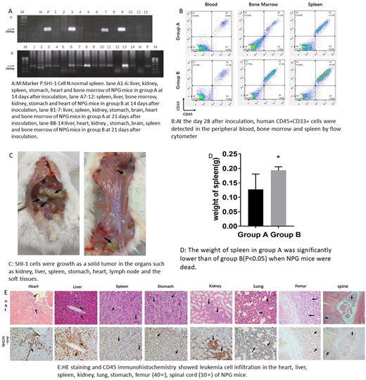
Although tumor cells are easily to growth in the bodies of immunodeficicent animals such as nude mice and NOD-SCID mice, it's hard for acute leukemia cells to grow in the bone marrow of nude mice or NOD-SCID mice even when mice receive extra immunosuppressive treatment such as splenectomy, cyclophosphamide and irradiation. This study aimed to establish a mice model with systemic leukemia using another highly immunodeficicent NPG mice without immunosuppressive treatment before inoculation. 5-week NPG mice were inoculated with 1x10⁷(Group A) or 5x10⁷ (Group B) SHI-1 cells (a cell line derived from a refractory acute monocytic leukemia patient) via tail vein. One NPG mice in each group was killed by ether randomly at the day 14, 21, 28 after inoculation, other NPG mice were observed the survival time. The leukemic cells engrafted in the NPG mice were detected by the following methods: the blast cells were detected by the blood smear and flow cytometer, the MLL-AF6 fuse gene of SHI-1 cells were detected by PCR amplification, the human CD45 positive cells infiltrated in the organs of NPG mice were detected by histopathological examination and immunohistochemistry. At the day 14 after inoculation with SHI-1 cells, fewer blasts cells were found in the smear of peripheral blood of group B; MLL-AF6 fuse gene could be amplified in the spleen of NPG mice in group A and in spleen and bone marrow in group B (Fig A); histopathological examination had shown that CD45 positive leukemia cell just infiltrated in spleen. At the day 21 after inoculation, more blasts were found in the smear of peripheral blood both in group A and B; MLL-AF6 fuse gene were amplified in the organs of NPG mice such as Spleen, liver, kidney, stomach, lung, heart and bone marrow(Fig A); 5.16% and 0.82% of CD45 and CD33 positive cells were detected in the peripheral blood of NPG mice in group A and B respectively; a green solid neoplasm were found in the kidney of NPG mice in group B, leukemia cells were found in the organ of heart, liver, spleen, stomach, kidney and lung in the NPG mice of both groups by histopathological examination. From the third week, the NPG mice presented anorexia, hunched posture, lethargy and weight loss. For the mice sacrificed in the day 28 after inoculation, the proportion of CD45 and CD33 positive cells in peripheral blood, bone marrow and spleen were 9.60%, 11.4% and 23.20% in group A and were 11.0%,37.80% and 60.5% in group B (Fig B). Green solid tumors were grown in many organs such as kidney, liver, spleen, stomach, heart, lymph node and the soft tissues in the NPG mice killed in day 28 after inoculation and the mice which were dead spontaneous (Fig C); When NPG mice were dead, the weight of spleen in group B is significantly higher than the weight of spleen in group A(P<0.05) (Fig D). The median survival time of NPG in group A and group B is 33 and 30 days respectively. Pathological examination and immunohistochemical staining had shown that leukemic cells could infiltrated to many of the organs of NPG mice and the grade of leukemia infiltration was positively correlated with cells numbers of inoculation and the survival time of NPG mice (Fig E). Altogether, SHI-1 cell could growth in the NPG mice without any pre-immunosuppressive treatment and formed a systemic leukemia in NPG mice as like in acute leukemia patients. This efficient and reproducible model may be a useful tool for the studies of the pathogenesis in acute monocytic leukemia.
No relevant conflicts of interest to declare.

This feature is available to Subscribers Only
Sign In or Create an Account Close Modal